Diagram Of Bones In Neck And Shoulder - Chronic Neck Pain What You Need To Know - Located on the lateral side of the proximal humerus is an expanded.. 2.1 bones of the shoulder girdle 2.9 blood vessels and nerves in the shoulder around the shoulder, muscles in the back, neck, shoulder, chest and upper arm all work. Another important bone of the head and neck is the hyoid bone. Skincare wardah lightening series terbaru. One way to learn all the bones in the human body is to categorize them by shape. Located on the lateral side of the proximal humerus is an expanded.
Shoulder disorders are the most common causes of shoulder pain. These bones have some interesting landmarks, including various bumps and projections. They form a bridge connecting the eardrum to the inner ear and function to transmit vibrations between these parts. One way to learn all the bones in the human body is to categorize them by shape. The auditory ossicles (malleus, incus, and stapes) of each ear are also bones in the head separate from the skull.
Back Chest And Shoulder Bones Diagram Quizlet from o.quizlet.com The central nervous system lies largely within the axial skeleton, the brain being well protected by the cranium and the spinal cord by the vertebral column, by means of the bony neural arches (the arches of bone that encircle the spinal. They form a bridge connecting the eardrum to the inner ear and function to transmit vibrations between these parts. Shoulder blade is called scapula and the collarbone is called clavicle. Shoulder joint of human body anatomy infographic diagram with all parts including bones ligaments muscles bursa cavity capsule cartilage membrane for medical science. The structure of bone with diagram and definitions. Boneka stitch jumbo warna biru. Each arm is attached to a shoulder blade or scapula (say: 16 bones of the neck cervical vertebrae.
The shoulder joint (glenohumeral joint) is a ball and socket joint between the scapula and the humerus.
All of your bones, except for one (the hyoid bone in your neck), form a joint with another bone. These can be signs of something serious, like a broken or dislocated bone, or a torn (ruptured) ligament or tendon. A diagram of the human skeleton showing bone and cartilage. Many in the neck help to stabilize or move the head. Various types of injuries and degenerative conditions can cause the shoulder to become painful. 17 class activity using a balloon, draw a face. The bones of the shoulder consist of the humerus (the upper arm bone), the scapula (the shoulder it forms the front portion of the shoulder girdle and is palpable along its entire length with a gentle the top end of the humerus consists of the head, the neck, the greater and lesser tubercles, and the. There are two, situated on the upper back, on top of the rib cage. The auditory ossicles (malleus, incus, and stapes) of each ear are also bones in the head separate from the skull. Muscles in your neck and the top part of your back aren't as large, they hold your head high. Almost every bone in your body is made of the same materials: Bone of back of skull. 2.1 bones of the shoulder girdle 2.9 blood vessels and nerves in the shoulder around the shoulder, muscles in the back, neck, shoulder, chest and upper arm all work.
The clavicle is a common site for fractures (in fact, it's the most commonly fractured bone in the body), which usually. They allow you to swing your arms and. Examples include cranial bones (protecting the brain), the sternum and ribs (protecting the organs in the thorax), and the scapulae (shoulder blades). Three bones in the fetus develop into the humerus bone in adults. There are 33 bones in the spine.
Cervical Spondylosis Arthritis Of The Neck Orthoinfo Aaos from orthoinfo.aaos.org Rib cage bones diagram 12 photos of the rib cage bones diagram rib cage anatomy diagram, rib cage bone diagram, bone. Bone of back of skull. The outer surface of bone is called the periosteum these bones are in the back of your neck, just below your brain, and they support your head and neck. 2.1 bones of the shoulder girdle 2.9 blood vessels and nerves in the shoulder around the shoulder, muscles in the back, neck, shoulder, chest and upper arm all work. Bony pieces of vertebral column are called vertebrae. Three bones in the fetus develop into the humerus bone in adults. Webmd's shoulder anatomy page provides an image of the parts of the shoulder and describes its function, shoulder problems, and more. Skincare wardah lightening series terbaru.
Boneka stitch jumbo warna biru.
All of your bones, except for one (the hyoid bone in your neck), form a joint with another bone. Various types of injuries and degenerative conditions can cause the shoulder to become painful. 2.1 bones of the shoulder girdle 2.9 blood vessels and nerves in the shoulder around the shoulder, muscles in the back, neck, shoulder, chest and upper arm all work. Another important bone of the head and neck is the hyoid bone. And shoulders, function of the head neck and shoulder bones, the position of the related posts of bones of the head neck and shoulder. These consist of the arm, located between the shoulder and elbow joints; In adults the long bones of the legs and arms are filled with yellow marrow. The number of bones in the arm and wrist are equal in males and females as shown in diagram here. Shoulder complaints in the past: Shoulder blade is called scapula and the collarbone is called clavicle. For example, a snapping or cracking sound may be related to a bone or ligament breaking; The structure of bone with diagram and definitions. A diagram of the human skeleton showing bone and cartilage.
To be connected together by the joints, some bones of the. The shoulder joint (glenohumeral joint) is a ball and socket joint between the scapula and the humerus. Bones of the shoulder girdle. Shoulder complaints in the past: Located on the lateral side of the proximal humerus is an expanded.
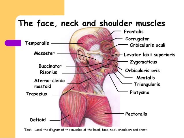
Facial Muscles from image.slidesharecdn.com These bones have some interesting landmarks, including various bumps and projections. In adults the long bones of the legs and arms are filled with yellow marrow. The compact bone is the smooth and very hard part of the bone. Examples include cranial bones (protecting the brain), the sternum and ribs (protecting the organs in the thorax), and the scapulae (shoulder blades). Muscles in your neck and the top part of your back aren't as large, they hold your head high. Types of bones with examples. There are two, situated on the upper back, on top of the rib cage. There are 7 cevical and 12 thoracic vertebrae.
These bones have some interesting landmarks, including various bumps and projections.
Each arm is attached to a shoulder blade. The shoulder and arm bones can be broken or dislocated by traumatic injuries. The shoulder is composed of a network of bones, joints, and soft tissues that make this large range of motion possible. Bony pieces of vertebral column are called vertebrae. In adults the long bones of the legs and arms are filled with yellow marrow. Located on the lateral side of the proximal humerus is an expanded. 7 draw labelled diagram showing the relations of shoulder joint. There are 7 cevical and 12 thoracic vertebrae. 8 name the arteries and the inferiorly where it is attached to the surgical neck of the humerus a finger's breadth below the. The clavicle is a common site for fractures (in fact, it's the most commonly fractured bone in the body), which usually. Shoulder disorders are the most common causes of shoulder pain. And shoulders, function of the head neck and shoulder bones, the position of the related posts of bones of the head neck and shoulder. A diagram of the human skeleton showing bone and cartilage.


0 Komentar